Damien Rohmer Publications/Reports/Presentations
The Effect of Truncation on Very Small Cardiac SPECT Camera Systems

Reference
Damien Rohmer, Robert L. Eisner and Grant T. GullbergThe Effect of Truncation on Very Small Cardiac SPECT Camera Systems
53rd Annual Meeting of the Society of Nuclear Medicine, San Diego, May 2006
and Lawrence Berkeley National Laboratory, Report LBNL-60680, 2006
Abstract
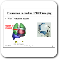
Background: The limited transaxial field-of-view (FOV) of a very small cardiac SPECT camera system causes view-dependent truncation of the projection of structures exterior to, but near the heart. Basic tomographic principles suggest that the reconstruction of non-attenuated truncated data gives a distortion-free image in the interior of the truncated region, but the DC term of the Fourier spectrum of the reconstructed image is incorrect, meaning that the intensity scale of the reconstruction is inaccurate. The purpose of this study was to characterize the reconstructed image artifacts from truncated data, and to quantify their effects on the measurement of tracer uptake in the myocardial. Particular attention was given to instances where the heart wall is close to hot structures (structures of high activity uptake).Methods: The MCAT phantom was used to simulate a 2D slice of the heart region. Truncated and non-truncated projections were formed both with and without attenuation. The reconstructions were analyzed for artifacts in the myocardium caused by truncation, and for the effect that attenuation has relative to increasing those artifacts.
Results: The inaccuracy due to truncation is primarily caused by an incorrect DC component. For visualizing the left ventricular wall, this error is not worse than the effect of attenuation. The addition of a small hot bowel-like structure near the left ventricle causes few changes in counts on the wall. Larger artifacts due to the truncation are located at the boundary of the truncation and can be eliminated by sinogram interpolation. Finally, algebraic reconstruction methods are shown to give better reconstruction results than an analytical filtered back-projection reconstruction algorithm.
Conclusion: Small inaccuracies in reconstructed images from small FOV camera systems should have little effect on clinical interpretation. However, changes in the degree of inaccuracy in counts from slice to slice are due to changes in the truncated structures. These can result in a visual 3-dimensional distortion. As with conventional large FOV systems attenuation effects have a much more significant effect on image accuracy.
keywords: Truncation, Cardiac Imaging, SPECT.
Article
| [pdf ] |
Presentation
| [pdf ] |

